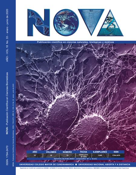
Hep-2 cells infected with eb’s from chlamydia trachomatis serovar 2 (vr-902b): research perspectives
Células hep-2 infectadas con eb’s de chlamydia trachomatis serovar 2 (vr-902b)
NOVA by http://www.unicolmayor.edu.co/publicaciones/index.php/nova is distributed under a license creative commons non comertial-atribution-withoutderive 4.0 international.
Furthermore, the authors keep their property intellectual rights over the articles.
Show authors biography
Chlamydia trachomatis (C. Trachomatis) is a Gram negative unmoving bacterium, characterized by being an obligate intracellular microorganism and having a reproductive cycle in which a metabolically inactive extracellular infectious form (elementary body - EB's) can be distinguished, and a non-non-cellular form. intracellular and active infectious (reticulated body - RB's). C trachomatis is characterized by causing infection in humans, is related to sexually transmitted diseases and eye infections, so it can lead to sequelae of interest if timely treatment is not given. The objective of this study was to optimize the infection model of C. trachomatis in HEp-2 cells with elementary bodies (EB’s) of C. trachomatis serovar L2. Initially, the conditions for the adequate growth of HEp-2 cells were established in time and with a confluence of 90%, to continue with the optimization of an infection protocol. The infection was confirmed from the staining with Giemsa allowing to evaluate morphological characteristics of both uninfected and infected HEp-2 cells and also of the elementary bodies of C. trachomatis. Finally, the infection was corroborated with the Direct Immunofluorescence technique that detects the C. trachomatis MOMP membrane protein. After the tests performed, the presence of nearby elementary bodies and within the cellular cytoplasm was evidenced, as well as vacuolated cells and cellular damage caused by the infection.
Article visits 351 | PDF visits 176
Downloads
Ramírez N Gloria, Vera A. Víctor J, Villamil J, Luis C. Cultivos Celulares, Elemento Fundamental para la Investigación. Revista Acovez. 2018;24(1).
2. Toolan HW. Transplantable human neoplasms maintained in cortisone-treated laboratory animals: H.S. No. 1; H.Ep. No. 1; H.Ep. No. 2; H.Ep. No. 3; and H.Emb.Rh. No. 1. Cancer research. 1954;14(9):660-6.
3. Instituto nacional de seguridad y salud en el trabajo. Chlamydia trachomatis- Fichas de agentes biológicos- DB-B-C.tr-16 España2016 [cited 2017]. Available from: https://www.insst.es/documents/94886/353495/Clamydia+trachomatis+2017.pdf/471a1569-928f-4c86-938b-9afd06ee360f?version=1.0.
4. Cardona-Arias JA, Gallego-Atehortúa LH, Ríos-Osorio LA. Infección por Chlamydia trachomatis en pacientes de una institución de salud de Bogotá y Medellín, 2012-2015. Revista chilena de infectología. 2016;33:513-8.
5. MARTÍNEZ T. MA. Diagnóstico microbiológico de Chlamydia trachomatis: Estado actual de un problema. Revista chilena de infectología. 2001;18:275-84.
6. ATCC. Chlamydia trachomatis (ATCC® VR-902B™) 2019 [cited 2017 8 abril 2017]. Available from: https://www.atcc.org/en/Products/Cells_and_Microorganisms/Viruses/Chlamydia_and_Rickettsia/VR-902B.aspx.
7. Jutinico Shubach AP, Mantilla Galindo A, Sánchez Mora RM. Regulación de la familia de proteínas BCLl-2 en células infectadas con Chlamydia Trachomatis. Nova. 2015;13:83-92.
8. Engstrom P, Bergstrom M, Alfaro AC, Syam Krishnan K, Bahnan W, Almqvist F, et al. Expansion of the Chlamydia trachomatis inclusion does not require bacterial replication. International journal of medical microbiology : IJMM. 2015;305(3):378-82.
9. Pajaniradje S, Mohankumar K, Pamidimukkala R, Subramanian S, Rajagopalan R. Antiproliferative and apoptotic effects of Sesbania grandiflora leaves in human cancer cells. BioMed research international. 2014;2014:474953.
10. Jutinico Shubach A, Malagón Garzón E, Sánchez Mora R. Cultivo de la línea celular HEp-2: doblaje poblacional y coloración con Giemsa Perspectivas para el estudio de la infección con Chlamydia trachomatis. Nova. 2013;11:23-33.
11. Nans A, Saibil HR, Hayward RD. Pathogen-host reorganization during Chlamydia invasion revealed by cryo-electron tomography. Cellular microbiology. 2014;16(10):1457-72.
12. da Cunha M, Pais SV, Bugalhao JN, Mota LJ. The Chlamydia trachomatis type III secretion substrates CT142, CT143, and CT144 are secreted into the lumen of the inclusion. PloS one. 2017;12(6):e0178856.
13. Derrick T, Last AR, Burr SE, Roberts CH, Nabicassa M, Cassama E, et al. Inverse relationship between microRNA-155 and -184 expression with increasing conjunctival inflammation during ocular Chlamydia trachomatis infection. BMC infectious diseases. 2016;16:60.
14. Richard Coico. Current Protocols in Microbiology. 2006 2019. In: Wiley Microbiology & Virology [Internet]. Available from: https://www.wiley.com/en-co/Current+Protocols+in+Microbiology-p-9780471729242.
15. Vicetti Miguel RD, Henschel KJ, Duenas Lopez FC, Quispe Calla NE, Cherpes TL. Fluorescent labeling reliably identifies Chlamydia trachomatis in living human endometrial cells and rapidly and accurately quantifies chlamydial inclusion forming units. Journal of microbiological methods. 2015;119:79-82.
16. Ibana JA, Sherchand SP, Fontanilla FL, Nagamatsu T, Schust DJ, Quayle AJ, et al. Chlamydia trachomatis-infected cells and uninfected-bystander cells exhibit diametrically opposed responses to interferon gamma. Scientific reports. 2018;8(1):8476.
17. Becker E, Hegemann JH. All subtypes of the Pmp adhesin family are implicated in chlamydial virulence and show species-specific function. MicrobiologyOpen. 2014;3(4):544-56.
18. Al-Zeer MA, Al-Younes HM, Lauster D, Abu Lubad M, Meyer TF. Autophagy restricts Chlamydia trachomatis growth in human macrophages via IFNG-inducible guanylate binding proteins. Autophagy. 2013;9(1):50-62.
19. Giakoumelou S, Wheelhouse N, Brown J, Wade J, Simitsidellis I, Gibson D, et al. Chlamydia trachomatis infection of human endometrial stromal cells induces defective decidualisation and chemokine release. Scientific reports. 2017;7(1):2001.
20. Soysa P, Jayarthne P, Ranathunga I. Water extract of Semecarpus parvifolia Thw. leaves inhibits cell proliferation and induces apoptosis on HEp-2 cells. BMC complementary and alternative medicine. 2018;18(1):78.
21. Cochrane M, Armitage CW, O'Meara CP, Beagley KW. Towards a Chlamydia trachomatis vaccine: how close are we? Future microbiology. 2010;5(12):1833-56.
22. Nogueira AT, Braun KM, Carabeo RA. Characterization of the Growth of Chlamydia trachomatis in In Vitro-Generated Stratified Epithelium. Frontiers in cellular and infection microbiology. 2017;7:438.
23. Carrera Páez LC, Pirajan Quintero ID, Urrea Suarez MC, Sanchez Mora RM, Gómez Jiménez M, Monroy Cano LA. Comparación del cultivo celular de HeLa y HEp-2: Perspectivas de estudios con Chlamydia trachomatis. Nova. 2015;13:17-29.
24. Gutiérrez DL, Sánchez Mora RM. Tratamientos alternativos de medicina tradicional para Chlamydia trachomatis , agente causal de una infección asintomática. Nova. 2018;16:65-74.
25. Sherrid AM, Hybiske K. Chlamydia trachomatis Cellular Exit Alters Interactions with Host Dendritic Cells. Infection and immunity. 2017;85(5).
26. Nguyen PH, Lutter EI, Hackstadt T. Chlamydia trachomatis inclusion membrane protein MrcA interacts with the inositol 1,4,5-trisphosphate receptor type 3 (ITPR3) to regulate extrusion formation. PLoS pathogens. 2018;14(3):e1006911.


